Fatemeh Momeni recently received the Best Poster Award at the iSFFF Conference in Washington, DC. Fatemeh’s poster highlighted her research on the retention behavior of different PFAS compounds in cyclical electrical field-flow fractionation.
Congratulations to Brady Goenner and Bahar Kazemi for successfully defending their dissertations!
Brady defended his dissertation entitled “Design Automation for 3D printed Microfluidics” last Wednesday, and Bahar defended her dissertation entitled “Implementation and Validation of a Programmable Modular Microfluidic Platform for Automated Metatranscriptomic Library Preparation” last Thursday.
We congratulate them on this important milestone and wish them continued success in their future careers.
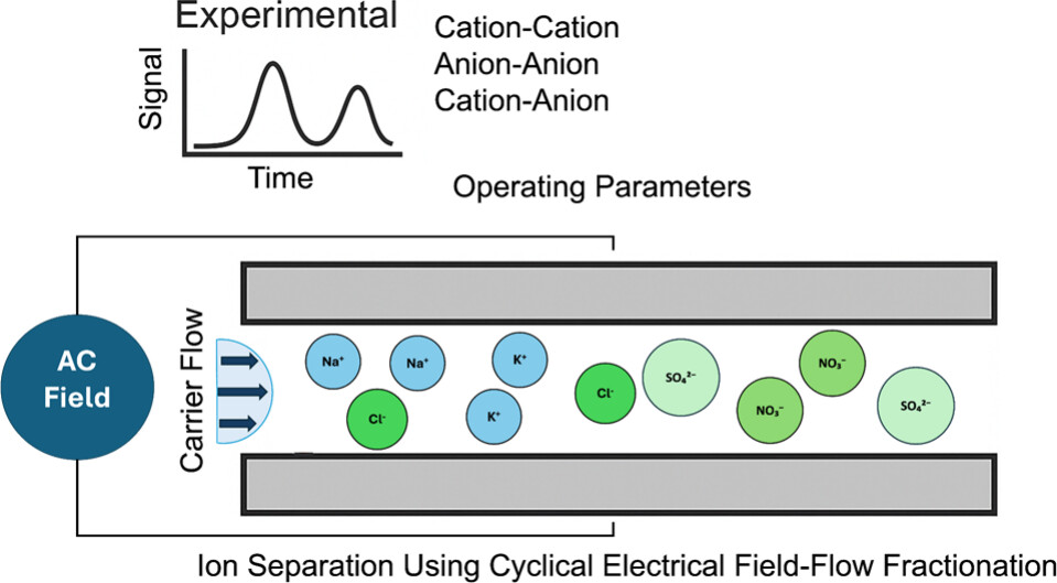
Mark Shad et al. published “Effect of Key Operating Parameters on the Retention and Separation of Ions Using Cyclical Electrical Field Flow Fractionation” in Analytical Chemistry. The full article can be found at https://doi.org/10.1021/acs.analchem.5c03277.
Graphical abstract.
Abstract: The analysis of inorganic ions is critical in the environmental, industrial, and medical fields, with ion chromatography (IC) being the most used method. While IC offers high accuracy and reproducibility, its high cost, complexity, and operational constraints necessitate the development of alternative methods. This study explores the application of CyElFFF, a variant of electrical field-flow fractionation, for ion retention and separation. The CyElFFF with channel dimensions of 25 μm height, 10 cm length, and 0.5 cm width was developed, incorporating a conductivity detector and CO2 suppressor. Parameters affecting retention time and separation resolution, including injection volume, ion concentration, and electric field conditions, were evaluated. Results showed enhanced ion retention and separation resolution at lower ion concentrations and optimized electric field settings in mode III. Separation of K+, Na+, NO3–, and SO42– was demonstrated with separation resolution improved by reducing frequency and flow rate. These findings establish CyElFFF as a promising approach for ion analysis.
Bruce Gale and Munawar Jawad received a UURF Interdisciplinary Research Initiative Track 1 seed grant from the University of Utah Vice President for Research (VPR) Office for a collaborative project focused on point-of-care screening for high-risk human papillomavirus (hrHPV). The project combines microfluidic engineering and molecular assay development to advance a low-cost, accessible diagnostic platform for cervical cancer screening. The work is carried out in collaboration with Natasha Pavlova and her team, with additional industry support from Patrick Manou.
In December, the Gale Group gathered to celebrate the end of the year with a Christmas party. Featuring a potluck, a white elephant gift exchange, and lively conversation, the event provided a great opportunity to relax outside the lab and strengthen connections.
Munawar Jawad et al. recently published “A disposable, passive microfluidic cartridge for point-of-care detection of antibodies in total capillary blood based on hemagglutination and machine-learning assisted interpretation” in RSC Advances.
Abstract: Point-of-care (PoC) detection of antibodies in blood enables rapid, on-site diagnosis. However, these devices often face challenges related to user variability due to the requirement of multiple manual operations. To address this issue, we designed and developed a disposable microfluidic device that requires minimal user input for rapid detection of SARS-CoV-2 antibodies (ABs) in total blood and antigens associated with blood types. Here, we present a passive pressure-driven pumping technique that rapidly mixes blood samples with reagents, delivering results within three minutes. The device requires 15 mL of capillary blood and can detect SARS-CoV-2 ABs across a concentration range of 0 to 60 mg mL−1. Additionally, we demonstrated the versatility of the microfluidic device by implementing blood typing functionality, highlighting its potential for broader serological testing applications. We also developed a support vector machine (SVM) algorithm as a proof-of-concept to demonstrate the potential application of machine learning (ML)-based analysis to complement visual interpretation of results. We evaluated the performance and predictive accuracy of the SVM model and compared it to human interpretations. The analysis showed that the SVM model achieved a statistically significant improvement in predicting varying degrees of agglutination when compared to human interpretation. This device addresses the need for a user-friendly, rapid COVID-19 AB testing solution and blood-typing assay and also provides a model for the future development of diagnostic devices that are integrated with ML models for improved diagnostic accuracy and accessibility in both clinical and non-clinical environments.