Fatemeh Momeni recently received the Best Poster Award at the iSFFF Conference in Washington, DC. Fatemeh’s poster highlighted her research on the retention behavior of different PFAS compounds in cyclical electrical field-flow fractionation.
Mark Shad et al. published “Effect of Key Operating Parameters on the Retention and Separation of Ions Using Cyclical Electrical Field Flow Fractionation” in Analytical Chemistry. The full article can be found at https://doi.org/10.1021/acs.analchem.5c03277.
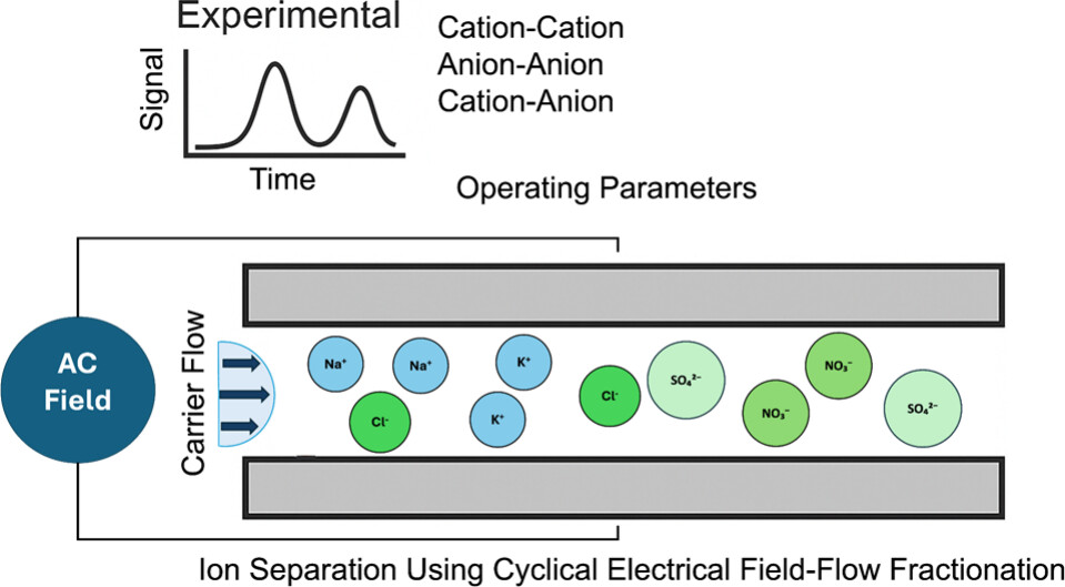
Graphical abstract.
Abstract: The analysis of inorganic ions is critical in the environmental, industrial, and medical fields, with ion chromatography (IC) being the most used method. While IC offers high accuracy and reproducibility, its high cost, complexity, and operational constraints necessitate the development of alternative methods. This study explores the application of CyElFFF, a variant of electrical field-flow fractionation, for ion retention and separation. The CyElFFF with channel dimensions of 25 μm height, 10 cm length, and 0.5 cm width was developed, incorporating a conductivity detector and CO2 suppressor. Parameters affecting retention time and separation resolution, including injection volume, ion concentration, and electric field conditions, were evaluated. Results showed enhanced ion retention and separation resolution at lower ion concentrations and optimized electric field settings in mode III. Separation of K+, Na+, NO3–, and SO42– was demonstrated with separation resolution improved by reducing frequency and flow rate. These findings establish CyElFFF as a promising approach for ion analysis.
Bruce Gale and Munawar Jawad received a UURF Interdisciplinary Research Initiative Track 1 seed grant from the University of Utah Vice President for Research (VPR) Office for a collaborative project focused on point-of-care screening for high-risk human papillomavirus (hrHPV). The project combines microfluidic engineering and molecular assay development to advance a low-cost, accessible diagnostic platform for cervical cancer screening. The work is carried out in collaboration with Natasha Pavlova and her team, with additional industry support from Patrick Manou.
Munawar Jawad et al. recently published “A disposable, passive microfluidic cartridge for point-of-care detection of antibodies in total capillary blood based on hemagglutination and machine-learning assisted interpretation” in RSC Advances.
Abstract: Point-of-care (PoC) detection of antibodies in blood enables rapid, on-site diagnosis. However, these devices often face challenges related to user variability due to the requirement of multiple manual operations. To address this issue, we designed and developed a disposable microfluidic device that requires minimal user input for rapid detection of SARS-CoV-2 antibodies (ABs) in total blood and antigens associated with blood types. Here, we present a passive pressure-driven pumping technique that rapidly mixes blood samples with reagents, delivering results within three minutes. The device requires 15 mL of capillary blood and can detect SARS-CoV-2 ABs across a concentration range of 0 to 60 mg mL−1. Additionally, we demonstrated the versatility of the microfluidic device by implementing blood typing functionality, highlighting its potential for broader serological testing applications. We also developed a support vector machine (SVM) algorithm as a proof-of-concept to demonstrate the potential application of machine learning (ML)-based analysis to complement visual interpretation of results. We evaluated the performance and predictive accuracy of the SVM model and compared it to human interpretations. The analysis showed that the SVM model achieved a statistically significant improvement in predicting varying degrees of agglutination when compared to human interpretation. This device addresses the need for a user-friendly, rapid COVID-19 AB testing solution and blood-typing assay and also provides a model for the future development of diagnostic devices that are integrated with ML models for improved diagnostic accuracy and accessibility in both clinical and non-clinical environments.
Brady Goenner et al. recently published “An open source platform to automate the design, verification, and manufacture of 3D printed microfluidic devices” in Scientific Reports.
Several barriers to widespread microfluidic adoption exist, including high initial fabrication costs and the labor-intensive development process. To address these barriers, a toolchain for the design, verification, and manufacturing of 3D printed microfluidic devices is presented. This work builds on existing electronic design automation (EDA) tools, yielding a toolchain that automatically lays out a microfluidic device from a library of components, simulates the device, and produces a 3D CAD file for manufacture via 3D printing. The process is validated by automatically designing and fabricating a calcium quantification assay. The full article can be found at https://doi.org/10.1038/s41598-025-15976-9.
Intracellular transformation and distribution of ZnO NPs in bacterial systems: CyElFFF-ICPMS fractograms showing time-dependent (0–24 h) change in intracellular ZnO NPs and Zn(II) ions in (A) S. fredii, and (B) B. subtilis; (C) Comparative biodistribution of ZnO NPs and Zn(II) ions in both bacterial species; Dissolution profiles of ZnO NPs to Zn(II) ions in (D) S. fredii and (E) B. subtilis; (H) Schematic diagram illustrating species-specific transformation pathway of ZnO NPs.
“Real-time monitoring of culture medium- and species-specific transformation of zinc oxide nanoparticles in bacterial systems using coupled CyElFFF-ICPMS,” authored by Weichen Zhao et al., was recently accepted in Microchemical Journal.
The article outlines the use of an online-coupled cyclical electrical field-flow fractionation and inductively coupled plasma mass spectrometry (CyElFFF-ICPMS) system for the quantification and characterization of the promising nanoagrochemicals of zinc oxide nanoparticles (ZnO NPs) and Zn(II) ions. Using the CyElFFF-ICPMS system, bacillus subtilis is found to stabilize ZnO NPs while sinorhizobium fredii promotes extracellular ZnO NP aggregation and dissolution. After 6 h, intracellular transformation significantly diverges. These findings demonstrate that the stability, transformation, and intracellular fate of ZnO NPs are highly governed by bacterial species-species interactions. The intracellular transformation and distribution of ZnO NPs in bacterial systems is displayed in the figure and the full article can be found at https://doi.org/10.1016/j.microc.2025.114926.
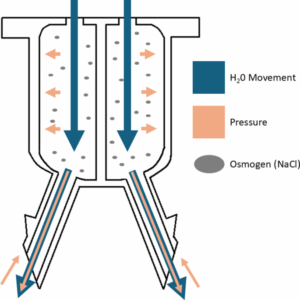
The working principle of the device. A diagram of water entering in through the semi-permeable membrane due to the high internal concentration of osmogen in the device, which generates a pressure gradient that propels the payload into the tissue, despite present intracranial pressure.
Ata Ullah and Jade Bookwalter et. al recently published “An Osmosis-driven 3D-printed brain implant for drug delivery” in Biomedical Microdevices.
To combat glioblastoma, a highly malignant brain tumor, several different drug-loaded devices have been developed to suppress tumor recurrence. However, these implants have limited effectiveness and often fail due to clogging, reflux, and limitations in intracranial implantation. Therefore, this article outlines the design, fabrication, and results of an osmosis-driven, 3D-printed brain implant. Featuring dual reservoirs, osmotic membranes, and precision-engineered needles, the implant achieves flow rates of 2.5±0.1 µl/Hr and diffusion distance up to 15.5±0.4 mm. A schematic of the working principle of the device is displayed in the figure and the full article can be found at https://doi.org/10.1007/s10544-025-00759-w.
Validation of the model using spherical polystyrene beads. Fluorescent images during a test (A) right before the outlets split, (B) along the spiral turns, (C) at the outlet split, and (D) captured outputs from each outlet after the test. No beads were collected in outlet 5.
“Application of Inertial Microfluidics for Isolation and Removal of Round Spermatids from a Spermatogenic Cell Sample to Assist In-Vitro Human Spermatogenesis” authored by Sabin Nepal, Joey Casalini, Alex Jafek, and Bruce Gale was recently published in Micromachines.
The article outlines the used of inertial microfluidics for isolating round spermatids from other germ cells and purifying spermatogenic cells as a way of improving in-vitro spermatogenesis to address male infertility. A custom PDMS microfluidic spiral channel for performing separation is designed, fabricated, and tested. The custom device does not experience clogging issues, a problem encountered in a commercially available spiral device. Additionally, the fabricated device achieves 86% purity in a single pass, an improvement over the 38% seen with STA-PUT – a method based on velocity sedimentation commonly used in this application. Validation results of the fabricated device are shown in the figure with the full article being found at https://doi.org/10.3390/mi16050500.
Schematic view of a waterborne parasitic protozoa detection system implementing microfluidic impedance flow cytometry.
Authored by Yunhao Peng, Bruce K. Gale, and Himanshu J. Sant, “Waterborne protozoan parasite detection using two-frequency impedance flow cytometry” was recently published in Analytical Methods.
A common cause of gastrointestinal diseases, waterborne parasitic protozoa are micron-sized parasites present in water sources. Therefore, the article outlines the development of a microfluidic water monitoring system based on impedance flow cytometry for the detection of these parasites. By utilizing parallel rather than coplanar electrodes, a limit detection of <0.1% volume ratio is achieved. Additionally, to improve sample discrimination, both a low and high frequency are applied simultaneously, making the method outlined in the article distinct from other proposed systems. A schematic of the monitoring system is displayed in the figure and the full journal article can be found at https://doi.org/10.1039/D5AY00184F.